Exploring Nanomagnetics: Detecting Muscle Movement with Precision
Technical Analysis | 04-07-2023 | By Robin Mitchell
Recently, researchers demonstrated a new textile that is able to detect slight movements in muscles and blood vessels when placed on the skin, utilising nanomagnetic materials. What challenges does monitoring sub-skin features present, what did the researchers demonstrate, and how could it be beneficial for future medical care?
What challenges does monitoring sub-skin features present?
Over millions of years, lifeforms (including humans) have developed an outer protective layer of skin and fat that helps to keep organs in place, prevent infection from pathogens, and provide adequate environmental shielding. But for all the benefits that such layers provide, modern medicine can often find them a nuisance. As organs and other internal body components are hidden, it is very difficult for doctors to directly observe tissue, and while this can be done via invasive procedures, the risk of infection and harm induced will often see such exploratory actions severely limited in scope and scale.
To get around this issue, medical technologies have developed numerous non-invasive techniques that allow for the internals of the human body to be imaged. One such technology is Magnetic Resonant Imaging (MRI) which takes advantage of how hydrogen atoms respond to strong magnetic fields. However, while MRI is certainly an advanced imaging technology, the high cost associated with MRI means that it is not often used unless essential (certainly in the case of the NHS).
Cheaper imaging systems, such as X-rays and CAT scans, take advantage of high-energy X-rays that can pass through certain tissue. The combination of advanced electronics and algorithms can further process internal images taken to provide doctors with plenty of diagnostic insight. However, the use of high-energy radiation introduces a cancer risk to patients. While this risk is very small, it still limits how often patients can be exposed to such systems.
Ultrasonic systems are another method used by doctors, and unlike MRI and X-rays, they are not only cheap but entirely harmless (so much so that they are used to image foetuses). The simple application of a gel combined with an ultrasonic transducer moved across the skin, sub-surface features can be identified, and this is particularly useful for finding damaged organs, inflammation, and some cancers. However, the poor penetration capabilities of ultrasonic waves combined with the poor resolution of generated images make them challenging to use.
Overall, most medical imaging technologies are either too expensive to deploy en-masse, induce a health risk, or provide poor-quality images.
In the quest for better imaging technologies, nanomagnetic materials have emerged as a promising solution. These materials, which are composed of tiny magnetic particles, have unique properties that make them ideal for medical imaging. According to a study published in the journal Matter, nanomagnetic materials can be used to create a textile that is sensitive enough to detect subtle changes under the skin, such as the movement of muscles and blood vessels.3
Researchers develop textile capable of detecting movement under the skin
Recently, researchers from the University of California published their findings on a newly developed textile that is capable of detecting subtle changes under the skin. The textile is made from a rubber patch impregnated with a nanomagnetic material and silver-coated yarn stitched into the patch via a sewing machine.
When the patch is placed over a piece of skin, any deformations in the patch result in a change in the magnetic field lines inside the patch. As changing magnetic fields induce currents, the tiny silver-coated yarn produces a voltage that is proportional to the amount of deformation that occurs.
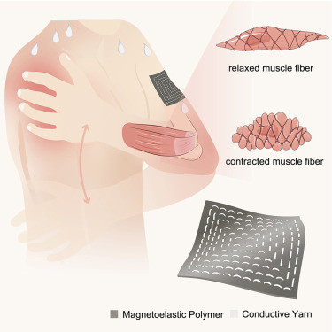
Due to the use of nanomagnetics in the rubber, it turned out that the rubber patch was highly sensitive to small changes in deformation and was demonstrated to produce clear, clean signals during blood vessel expansion and individual muscle movement. According to the researchers, the patch was sensitive enough to be able to distinguish between different muscle groups in test subjects.
To demonstrate the survivability of the sensor, the researchers also doused the patch in water to test how real-world scenarios (such as heavy sweating) would affect the patch. The result of numerous tests showed minimal impact on the patch, indicating that the patch is not only waterproof but also able to withstand numerous deformation cycles.
"Our work shows that nanomagnetic materials can be used to create a highly sensitive, flexible sensor that can detect subtle changes in the body," said Professor Zhenan Bao, the K.K. Lee Professor of Chemical Engineering at Stanford, who was not involved in the study. "This could open up new possibilities for non-invasive medical imaging and wearable health monitoring devices."1
How could this new patch help medical diagnosis?
The ability to detect subsurface movements with accuracy introduces multiple potential benefits, especially to wearable medical devices.
The first major use that such a sensor could introduce is the ability to measure blood pressure without the need for a constrictive device. Traditionally, taking blood pressure requires blood flow in an arm to be temporarily halted, and while this is perfectly adequate for routine testing via a medical practitioner, it is far from practical for everyday use. Instead, the newly developed patch could detect the subtle changes caused by veins and correlate the amount of deformation with pressure (such that less deformation implies lower blood pressure).
The second major use that this sensor could provide is for therapy and rehabilitation. By placing multiple sensors across various muscles, it is possible to identify which specific muscle groups are responding incorrectly. From there, medical practitioners can provide improved care that targets these muscle groups, thereby helping those going through rehabilitation.
Overall, what the researchers have developed is exciting, especially when considering that the sensor is extremely cheap to produce. How quickly it can be turned into something practical is still yet to be seen, but there is a good chance that nanomagnetic materials will find their way into wearable medical devices.
As the researchers noted in their study, "The development of this nanomagnetic textile is a significant step forward in the field of medical imaging. It offers a non-invasive, cost-effective solution that could revolutionise how we monitor and diagnose health conditions."3
"Our device is very sensitive to biomechanical pressure," says senior author Jun Chen of the Department of Engineering, University of California, Los Angeles. "The device converts muscle activities into quantifiable, high-fidelity electrical signals that are sent wirelessly to phone apps. This demonstrates the potential for personalised physical therapies and improving the rehabilitation of muscle injuries."1
To understand how the nanomagnetic textile works, it's important to first understand the properties of nanomagnetic materials. These materials are composed of tiny magnetic particles, each with a size on the order of nanometres. When these particles are placed in a magnetic field, they align themselves with the field, creating a uniform magnetic field within the material. This property is what allows the textile to detect changes in the body.
The textile itself is made from a rubber patch impregnated with the nanomagnetic material. Silver-coated yarn is then stitched into the patch, creating a network of conductive threads. When the patch is placed on the skin, any movements or deformations in the skin cause the magnetic field within the patch to change. This change in the magnetic field induces a current in the silver-coated yarn, which produces a voltage that is proportional to the amount of deformation. This voltage can then be measured and analysed to detect subtle movements in muscles and blood vessels.3
Reference Section:
- Eurekalert.org. (2023). Researchers develop textile capable ofdetecting movement under the skin. https://www.eurekalert.org/news-releases/993429
- Eurekalert.org. (2023). Multimedia associated with the research. https://www.eurekalert.org/multimedia/989508
- Cell.com. (2023). Full text of the research article. https://www.cell.com/matter/fulltext/S2590-2385(23)00294-1