Silicon Nanotechnology in Disposable Ultrasound Patches
Insights | 16-09-2025 | By Liam Critchley
Key Things to Know:
- Traditional ultrasound wearables rely on rigid, lead-based piezoelectric materials that face regulatory and comfort challenges.
- Capacitive micromachined ultrasonic transducers (CMUTs) provide a lead-free, flexible, and cost-effective alternative.
- Silicon nanocolumn CMUT (snCMUT) patches demonstrate high-resolution imaging, thin design, and scalability for disposable medical use.
- Clinical studies confirm CMUT-based patches can accurately monitor vascular dynamics and support long-term cardiovascular health monitoring.
Wearable technology has continued to grow for many years in the medical sector. A lot of wearable technology has been spearheaded by flexible substrate materials and ultra-thin materials, often with nanomaterials used as the active components. While both organic and inorganic nanomaterials have been used to create many wearable devices, ultrasound wearables have relied on bulkier inorganic piezoelectric materials to achieve high performance. However, the rigidity of these materials, combined with their environmental impact, as many rely on lead-based piezoelectric materials, has made it difficult to create many medical-grade ultrasound wearables.
There is an interest in creating new ultrasound wearables, as ultrasound is a widely used non-invasive medical imaging approach. There have been a lot of advancements in wearable ultrasonic transducer arrays that are now helping clinicians to monitor blood flow in different regions of the body, image cardiovascular structures, and for mapping tissues. Having more effective wearable ultrasonic devices that can adhere to more parts of the body would make ultrasound wearables a lot more valuable across different medical imaging applications.
Wearable Ultrasound Device Issues
Despite the interest in wearable ultrasound devices, there have been a number of material limitations. The first limitation is that the conventional piezoelectric material often used contains lead that violates the Restriction of Hazardous Substances Directive (RoHS) in the EU, making its clinical feasibility very difficult for many locations.
The second main issue is that piezoelectric materials require thick backing layers to improve ultrasound propagation, reduce pulse duration, and dampen vibrations. While this improves device performance, they are usually too thick to make devices that flex sufficiently for wearable devices and can be uncomfortable to wear. If the thickness of the backing layers is reduced, it leads to a poor resolution due to the reverberation of the transducers, which makes them unusable. It is also very difficult to assemble rigid piezoelectric materials with flexible electrodes, leading to oversized devices that account for the mechanical mismatch, which also makes the device uncomfortable for the patient.
While not a technical challenge, the cost of many ultrasound transducer arrays is much higher than is feasible for clinical settings. To promote good hygiene and infection control, any applied patch, including ultrasound patches, should be a one-use application that can be disposed of after use. So, to be feasible for use in hospitals, ultrasound patches would have to be commercially feasible as well as technologically capable.
Capacitive Micromachined Ultrasonic Transducers (CMUTs) Offers an Alternative Approach
Researchers have now taken to using capacitive micromachined ultrasonic transducers (CMUTs) as a lead-free approach to traditional piezoelectric material-based ultrasound transducers. CMUTs can avoid regulatory issues related to lead for medical imaging applications. They are also based on silicon wafer semiconductor fabrication technology, so they have the potential to be fabricated with existing batch fabrication lines in the industry to reduce the costs to levels suitable for the medical sector. Overall, CMUTs also have a better sensitivity, resolution, signal-to-noise ratio, and bandwidth compared to traditional piezoelectric ultrasound patches. Unlike piezoelectric materials that can rise in temperature over time, CMUTs also avoid any increase in temperature during operation, which makes them suitable for long-term medical monitoring.
Clinical Relevance of CMUT Technology
In clinical research, CMUT technology has shown promise for monitoring vascular dynamics continuously. According to Lee et al. (Nature Communications, 2025), the integration of CMUTs into flexible patches allows high-resolution imaging with minimal thermal variation, a critical factor for long-duration diagnostic use. This aligns with broader clinical goals of creating lead-free, biocompatible devices that conform to patient safety standards.
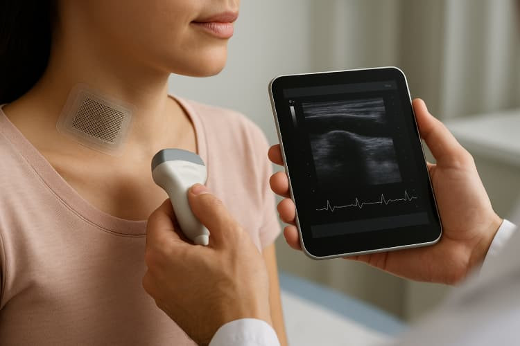
Using semiconductor fabrication and micromachining methods, researchers have now developed a skin-conformal silicon nanocolumn CMUT (snCMUT) array that can be integrated into disposable and wearable ultrasound patches for real-time ultrasonic imaging. The patches were very flexible, had a high-pressure output, a high transmission efficiency of 220 kPa/V, a low power consumption, and could be fabricated for under $20 each.
Scalable Manufacturing and Healthcare Integration
The affordability of the patches highlights their scalability for healthcare systems that require cost-effective disposable devices. In addition to low-cost production, the silicon nanocolumn CMUT design demonstrated transmission stability during extended operation, which is important for real-time monitoring in point-of-care settings. Such developments contribute to advancing medical imaging accessibility while maintaining compliance with regulatory frameworks like the EU RoHS directive.
The ultrasound patch was composed of individual CMUT elements separated by elastomer-filled trenches, all on top of a flexible printed circuit board (PCB). The PCB was then encapsulated with polydimethylsiloxane (PDMS). The silicon nanocolumns acted as springs beneath the moving plate of the snCMUT to increase the displacement and transmission efficiency. The snCMUT array could therefore flex above 1 mm radius of curvature and stretch up to 140% with degradation. The fully packaged snCMUT had a thickness of around 900 microns, enabling very thin monitoring patches to be built. To showcase the capabilities of the ultra-thin disposable patches, the researchers used them to study the carotid artery from both sides of the neck in real-time.
The study also confirmed that the thin profile of the snCMUT array enabled precise arterial wall visualisation, which is not always achievable with bulkier piezoelectric-based systems. The flexibility of the patch structure, capable of stretching up to 140%, ensures consistent skin contact during patient movement, thereby reducing imaging artefacts. These characteristics strengthen CMUT-based wearables as candidates for long-term cardiovascular monitoring and early disease detection.
Using the Patches to Study the Carotid Artery
The researchers used the patches to continuously monitor the carotid artery, obtaining key information about the blood pressure pulse waveforms, including the maximal slope, systolic peak, dicrotic notch, and diastolic peak. To perform the ultrasonic imaging, the researchers placed two disposable snCMUT patches on either side of the neck. The patches conformed to the neck and were tested on 9 healthy volunteers to monitor the left and right carotid arteries using M-mode ultrasound imaging. The high transmission efficiency of the patches enabled the carotid arteries, jugular vein, and sternocleidomastoid muscle to be easily distinguished using ultrasound.
The patch-enabled imaging allowed the researchers to look at the contraction and expansion of the carotid artery by measuring the positional changes of the anterior and posterior walls of the artery. This allowed the blood pressure changes to be tracked over time by converting the distance between the arterial walls into blood pressure pulse waves and comparing it with known values from a clinical-grade tonometer. The results from the patches showed a 96.5% agreement with the tonometer, showing a high accuracy.
While healthy volunteers don’t show much difference on either side of the carotid artery, placing two patches on either side of the carotid artery allows carotid artery asymmetry to be identified. When a patient exhibits carotid asymmetrical arterial stiffness, it can be an indicator of atherosclerosis, allowing more in-depth tests to be performed to confirm. Additionally, placing patches on either side of the neck allows conditions such as hypertension to be diagnosed, which can be missed if imaged from one side only.
Even though the patches operate at a very low voltage compared to other ultrasonic patches, the snCMUT patches obtained more precise ultrasound images than previously reported wearable ultrasound imagers, which can require voltage amplification up to 200 V. The flexible and disposable patches offer a low-cost and effective approach to wearable ultrasound monitoring that promotes patient comfort. The researchers have confirmed that while this study focused on creating the patches and imaging the carotid artery, more work is currently being undertaken to use the patches for imaging various parts of the body in real-time.
Reference: